The navicular bone is one of the 26 bones in the human foot. It’s important for connecting the ankle to the lower bones in our feet and helps form the arch that enables us to walk well. It is prone to stress fractures, especially by athletes while kicking, sprinting, twisting, or falling.
Anatomy
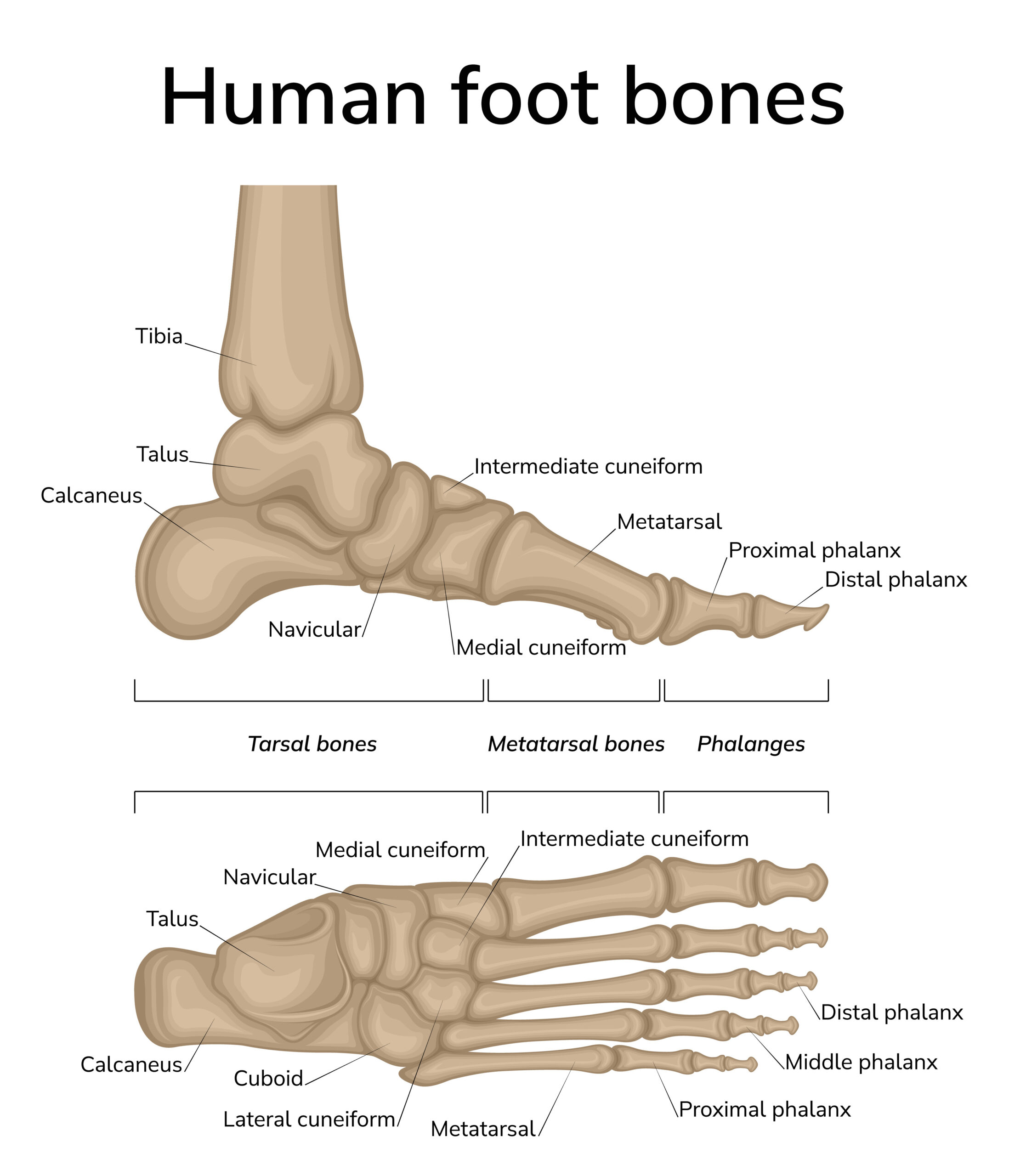
The human foot contains 26 bones and 33 individual joints. The navicular is a boat-shaped bone located in the top inner side of the foot, just above the transverse. It helps connect the talus, or ankle bone, to the cuneiform bones of the foot.
Function
Although small, the navicular bone serves important functions. Several ligaments and a tendon connect to the navicular bone, which stabilizes our ankle and arch. This allows us to walk efficiently.
Associated Conditions
Conditions that affect the navicular bone include fractures, Kohler disease, Mueller-Weiss syndrome, and accessory navicular syndrome.
Fractures
Acute fractures of the navicular bone can occur with injury. Stress fractures can occur from repeated stress.
Athletes commonly fracture the navicular bone while kicking, twisting, or sprinting. Pain and change in how you walk are common with fractures. Among track athletes, navicular stress fractures are one of the most common causes of stress fractures.
Treatment can be conservative or surgical, depending on the severity of the fracture. Conservative treatment includes casting to allow the bone to heal. Surgery can be required to place screws that will hold the bone in place along with a cast to allow time for healing.
Kohler Disease
Kohler disease is an avascular necrosis or osteonecrosis disease, which means there is a death of the bone tissue from lack of blood supply needed to keep bones healthy. Kohler disease is most commonly seen in young children between the ages of 4 and 7. It is more common in boys.
The navicular bone is calcified in children when they are around 3 years old. As children grow, their increasing weight makes the navicular bone susceptible to compression by other bones in the foot. This compression—along with the lack of blood supply to the navicular bone—increases the chances of Kohler disease.
Usual symptoms are tenderness and pain in the middle of the foot. Swelling may also be present.
This disease is self-limited and usually corrects itself as children mature. Anti-inflammatory medications and immobilization with a short walking cast are the recommended treatment to alleviate pain.
Muller-Weiss Syndrome
Muller-Weiss syndrome (MWS), also known as Brailsford disease, is an osteonecrosis disease, but it occurs in middle-aged adults. MWS is more frequently seen in women.
MWS can arise spontaneously, although some believe it is caused by trauma, bone migration, or could be due to congenital causes.
Conservation treatment of anti-inflammatories and immobilization are typically successful in treating pain. Surgical alternatives are available, if needed, to alleviate pain and to restore the arch.
Accessory Navicular Syndrome
Accessory navicular syndrome is the result of an extra bone or piece of cartilage alongside the navicular bone. This is a congenital condition and is present at birth. It occurs in 4 to 14% of the population.
Most people who have accessory navicular syndrome do not know it exists unless the extra bone causes a problem.
Trauma, foot or ankle sprain, and chronic irritation from footwear rubbing on this extra bone can cause pain.
Having flat feet, or fallen arches can also cause strain on the tendon that connects to the bone, which can cause accessory navicular syndrome to worsen and create more inflammation and pain.
Symptoms of accessory navicular syndrome include:
- Pain or throbbing in the midfoot and arch—typically during/after activity
- Visible prominence or bump on the inner side of the foot, above the arch
- Redness and swelling of the bony prominence
In adolescence, cartilage calcifies, which turns into bone. Often it is during this process when symptoms of accessory navicular syndrome appear. Some people don’t experience symptoms until later in life.
History of pain, examination, and x-rays can confirm the diagnosis of accessory navicular syndrome.
Ice to reduce swelling, anti-inflammatory medications, physical therapy to strengthen muscles and decrease inflammation, as well as immobilization in a cast may be used to treat symptoms. Another option for treatment is a custom orthotic device, which is inserted into the shoe to provide arch support.
If symptoms reappear after conservative treatment, surgery to remove the extra navicular bone may be needed.
Treatment
Recovery from navicular bone associated conditions typically include conservative treatment with immobilization and anti-inflammatories. Podiatry input and foot orthoses are often utilized to help prevent reinjury.
#stepandstridepodiatry #respod
Call us today to resolve your foot pain.
0800 473 776
(09) 212 9612